Virtual lab: Australopithecus afarensis knee joint
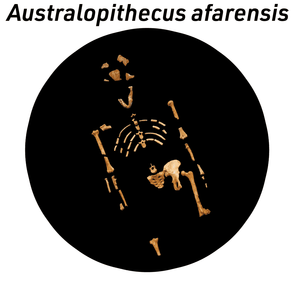
The largest sample of cranial material of Australopithecus afarensis comes from Hadar, Ethiopia. Fossils of Au. afarensis from this site range in age from 3.4 million to 2.9 million years ago. One of the earliest fossil hominin specimens found at Hadar was from the AL 129 locality, where the fragmentary femur and tibia of an ancient individual were found.

This virtual lab includes models of the distal portion of the femur and proximal tibia from a single individual of Au. afarensis, designated as AL 129-1. These two fragments show the form of the knee joint in this species.
The form of the knee joint in humans reflects our habitual bipedal locomotion. If you place the condyles of a human femur flat on a table surface, the shaft of the bone will have an angle. In humans, the shaft angles outward from the knee toward the hip. This direction of the angle is known as valgus, so it is often said that humans have a valgus knee. This angulation places the center of mass of the body more directly over the knee joint, when an individual is standing on one leg or taking a walking step. Other living primates do not have a valgus knee.
The condyles of the human femur are also shaped in a way that helps stabilize the knee joint while walking and running. In particular, from the lateral condyle running forward is a prominent ridge of bone that helps to keep the patella from dislocating laterally.
The condyles of the human tibia are more symmetrically shaped than those in the chimpanzee femur, and in particular the lateral tibial condyle is concave in humans, while it is somewhat convex in chimpanzees. The chimpanzee knee joint supports a greater range of reaction forces than human knees. Humans have knees that help to resist injury and transmit force in an extended leg, one-legged stance and gait.
As you inspect the Au. afarensis knee joint, make note of the characteristics that reflect bipedal walking and running in humans compared to chimpanzees.
Materials in this lab
- Original Au. afarensis fossil material from Hadar is curated at the National Museum of Ethiopia, Addis Ababa. The anatomical models of the AL 129-1 distal femur and proximal tibia in this virtual lab are based on measurements of casts of these specimens.
- The human and chimpanzee models in this lab are based on data from osteological specimens in the Biological Anthropology collection at UW-Madison.
Back to full list of virtual labs