
Virtual labs in Biological Anthropology
The COVID-19 crisis has been challenging for many instructors in biological anthropology. Our courses introduce students to the human skeleton, the anatomical diversity of non-human primates, and the fossil record of human evolution. In our teaching labs, students encounter real objects: human and non-human primate skeletal material and casts of fossils. Many students learn better from seeing and touching these objects than they can learn from lectures or reading. So being out of the lab is tough.
I've been teaching online since 2013. Until now I've never found a very satisfying way to present laboratory materials in a virtual 3D setting. Biological anthropology is a comparative science, and that means students need to be able to compare materials with each other. There are some great online resources with 3D data. But using 3D models usually requires students to download and learn special software, and none of these software packages make it easy to set up comparisons that show multiple objects in the same orientation and scale.
Once the crisis began, I started working to find a way to overcome the challenges of virtual labs. I found a solution based on WebGL and Javascript, which will work in most browsers without downloading special software. I've done a lot of testing, and have been able to run the labs in my course this semester with 560 students. I am sharing some of these labs here.
- The virtual labs work on phones, on iPads and other tablets, and on laptops and desktops using modern browsers. I find that they do not work on my oldest (2010-2013) iPads or older browsers.
- Just as with any process of reproducing or depicting skeletal remains, it is important to understand the limitations of this format. I've described the process and limitations in some detail on the Virtual Lab Frequently Asked Questions page.
- These virtual labs include anatomical models and not scans. In many cases, the creation of the model begins with a 3D surface scan of a bone or fossil. In others, the process begins from anatomical models built by artists. These are learning aids and are not suitable for research measurements.
- The virtual labs do not include models that can be extracted or printed. For those interested in 3D printable models, I have included links to resources in each virtual lab.
If you are an instructor and are interested in building some of these virtual labs into your course, please reach out to me. I do have self-contained virtual labs that I deploy on Canvas for my own students, which may be a better option for some courses than external links to my lab website. I also have some virtual labs that I am not releasing yet to the public. If these would be useful for your courses, I would love to hear from you.
My laboratory is sharing these low-resolution virtual laboratories exclusively for educational purposes. These virtual labs are available under a Creative Commons-Attribution-Noncommercial (CC-BY-NC) license. The construction of the virtual labs requires a low poly-count 3D representation of skeletal material that is not useful for printing, research, or commercial reproduction. My commitment with all educational materials is for them to work well on a phone, bringing them into reach for students worldwide outside of university settings. My lab's work with these materials is made possible by the scientific collaboration of many institutions around the world. The sources for anatomical models and data are listed in each virtual lab. In addition, I have linked sources for high-resolution 3D data and anatomical models where they exist for each specimen.
Human skeletal labs
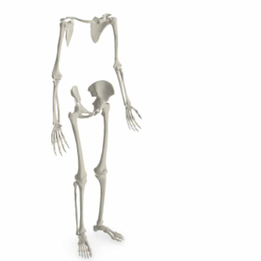
Regions of the skeleton
Introducing the cranium and postcranial skeleton, axial and appendicular skeleton, pelvis, vertebral column, and shoulder girdle.
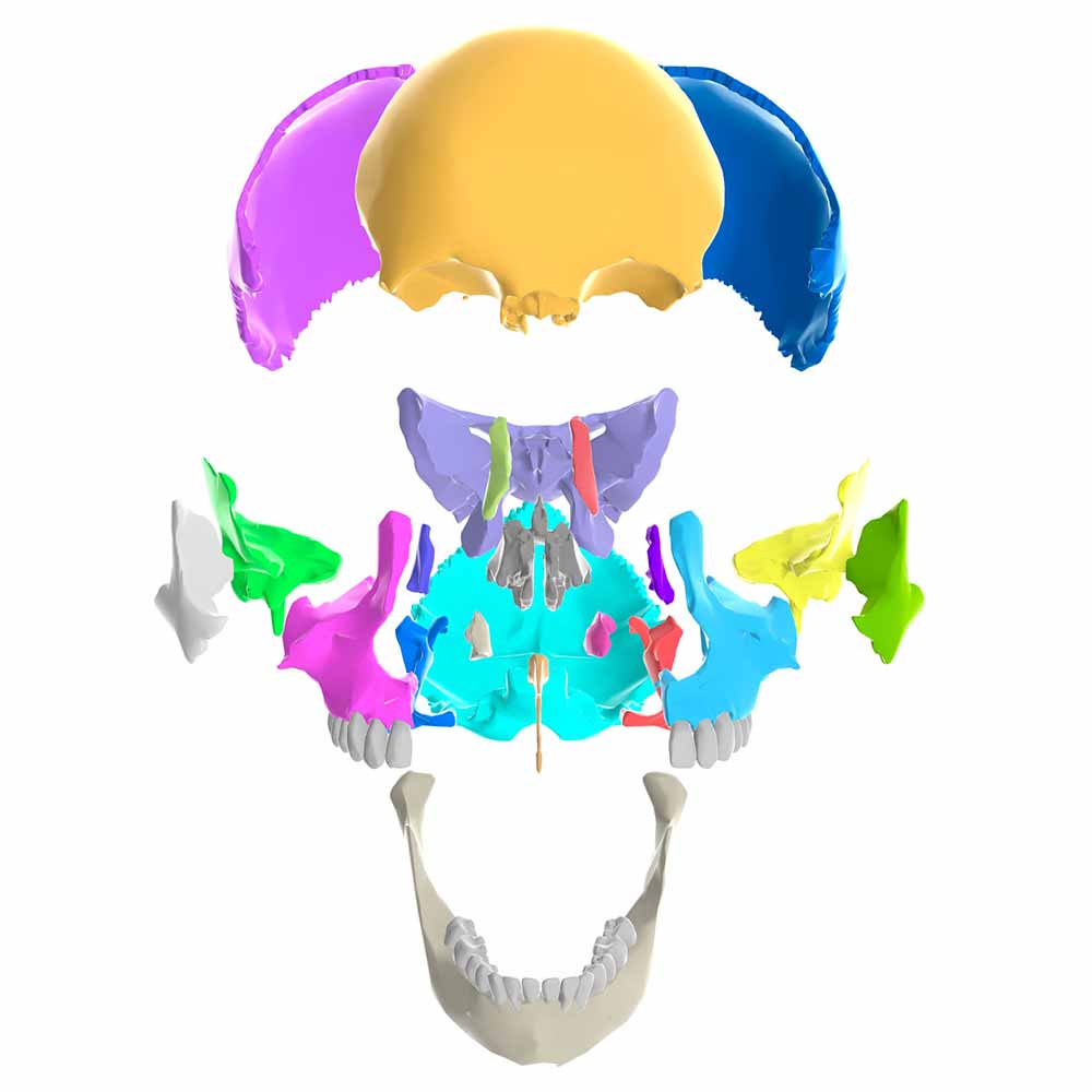
Regions of the cranium
Delineating the cranial vault, face, and mandible, with individual bones labeled and colored.
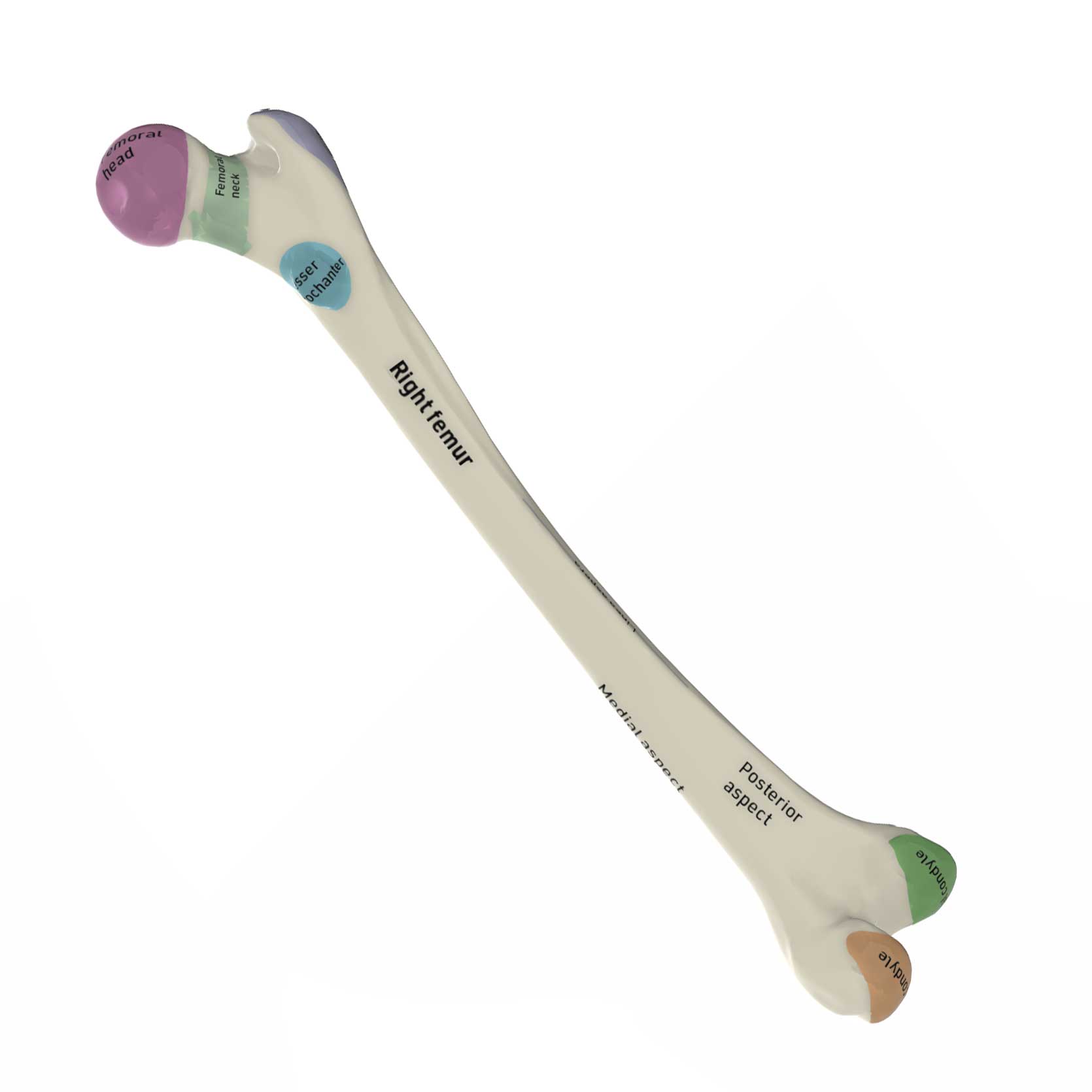
Morphology of the femur
Illustrating features of the femur in humans with labels.
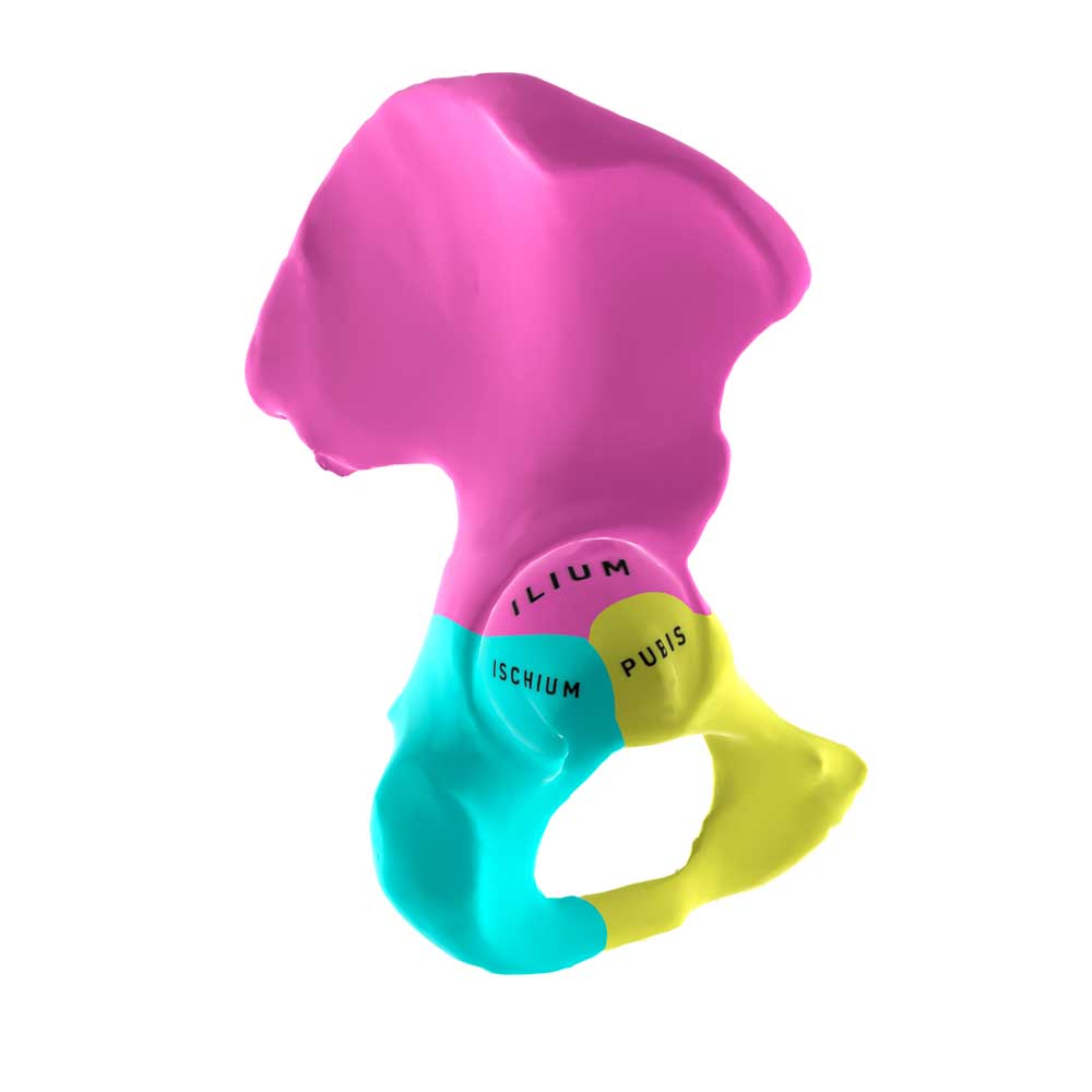
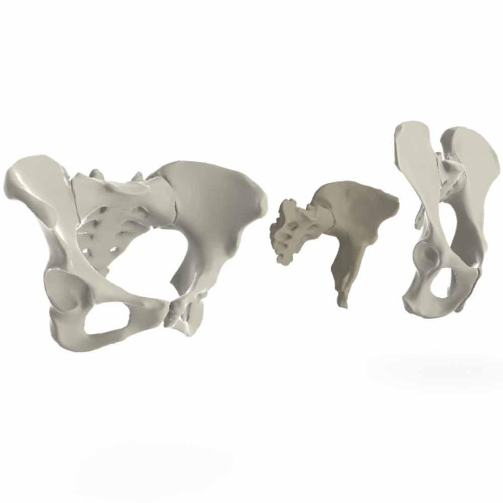
Morphology of the pelvis
A tour of the ilium, ischium, pubis, and sacrum.
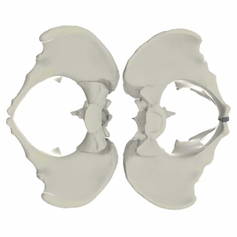
Pelvic sexual dimorphism
Comparing the pelvic morphology associated with male and female skeletal sex.
Australopithecus
Pelvic anatomy of Au. afarensis
The iconic AL 288-1 (Lucy) pelvic remains compared with human and chimpanzee pelves.
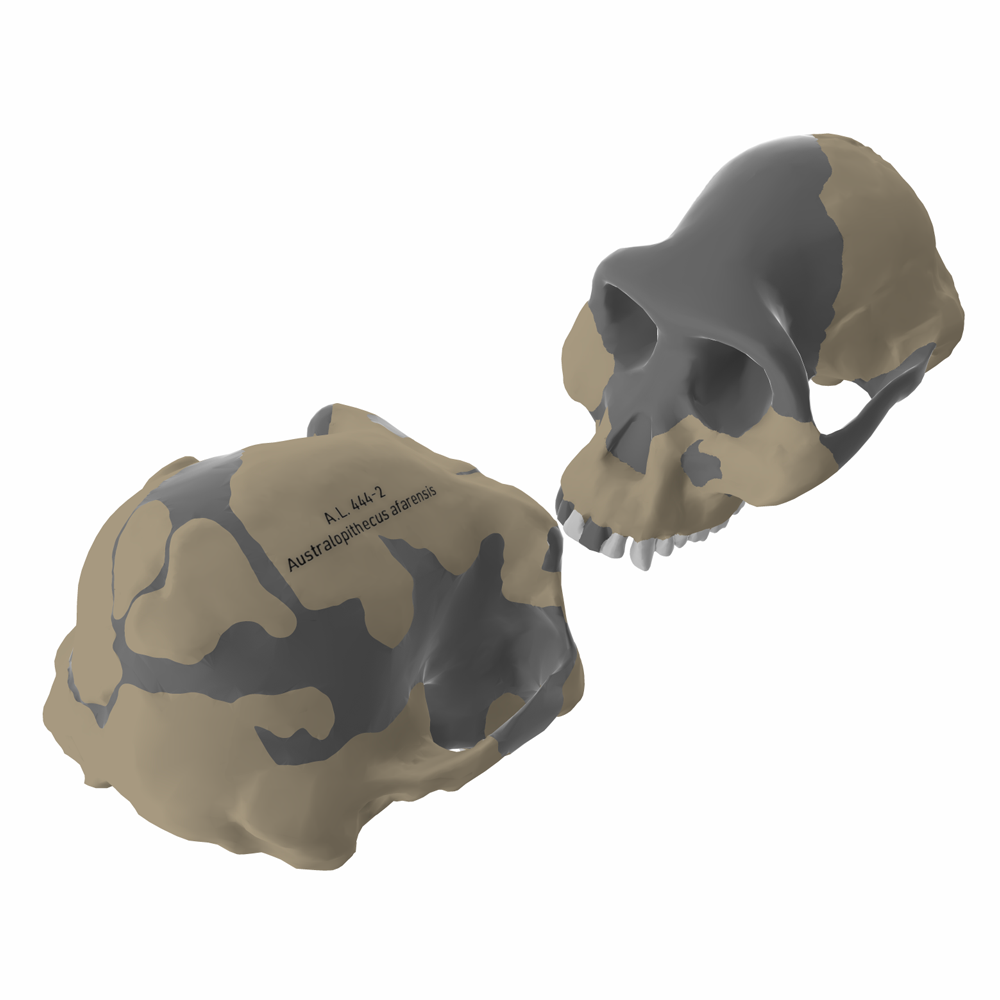
Australopithecus afarensis crania
The composite reconstruction from the AL 333 site, together with AL 444-2.
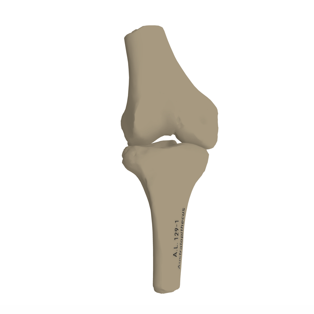
Australopithecus afarensis knee joint
The AL 129-1 femur and tibia, illustrating the valgus hominin knee joint.
Cranial anatomy of Au. africanus
A look at the iconic Sts 5 skull, with two other Sterkfontein cranial fossils.
The Taung skull
Introducing the first discovery of Australopithecus africanus in comparison to juvenile human and chimpanzee crania.
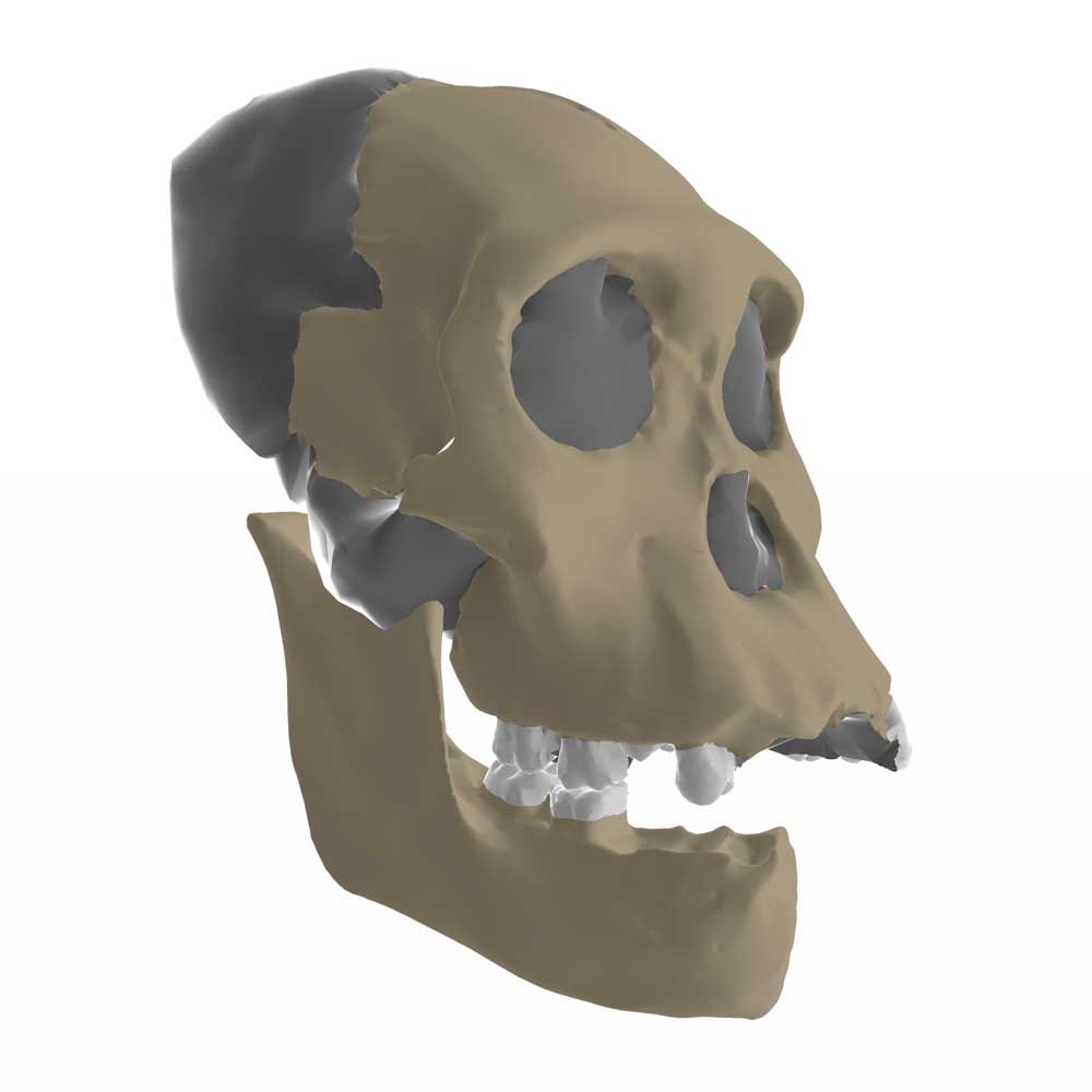
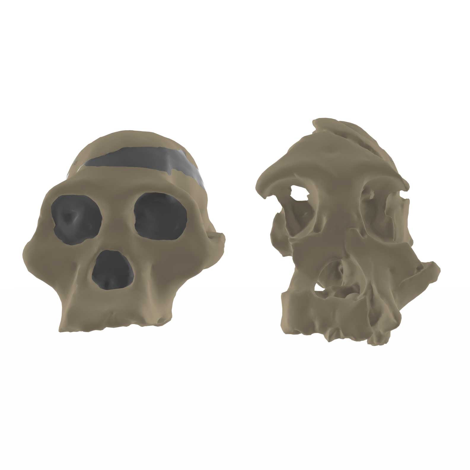
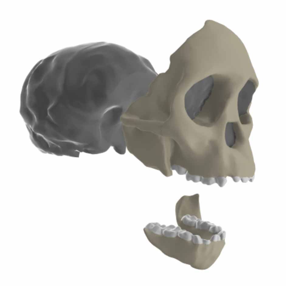
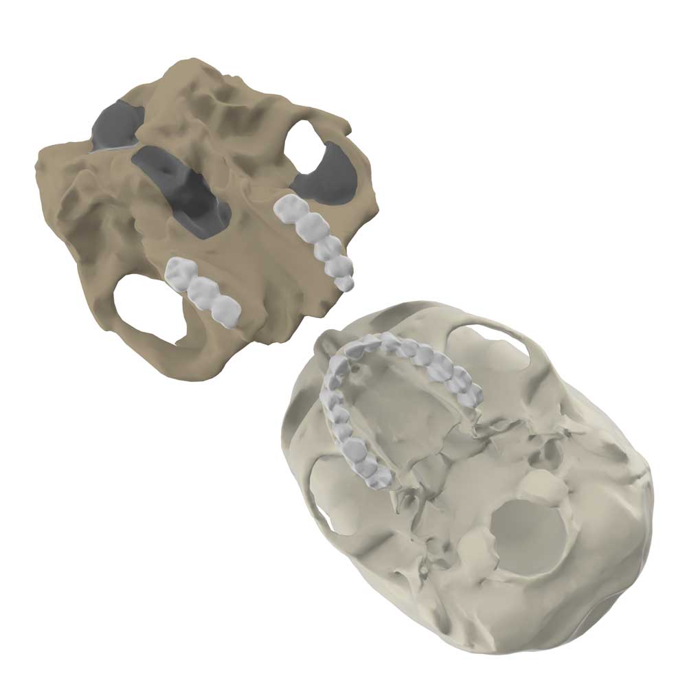
Australopithecus sediba with MH1
A comparison of the MH1 juvenile cranium with D2700 from Dmanisi and Sts 5.
Paranthropus
Paranthropus robustus crania
TM 1517 from Kromdraai and SK 48 from Swartkrans, compared to Sts 5 and a modern human.
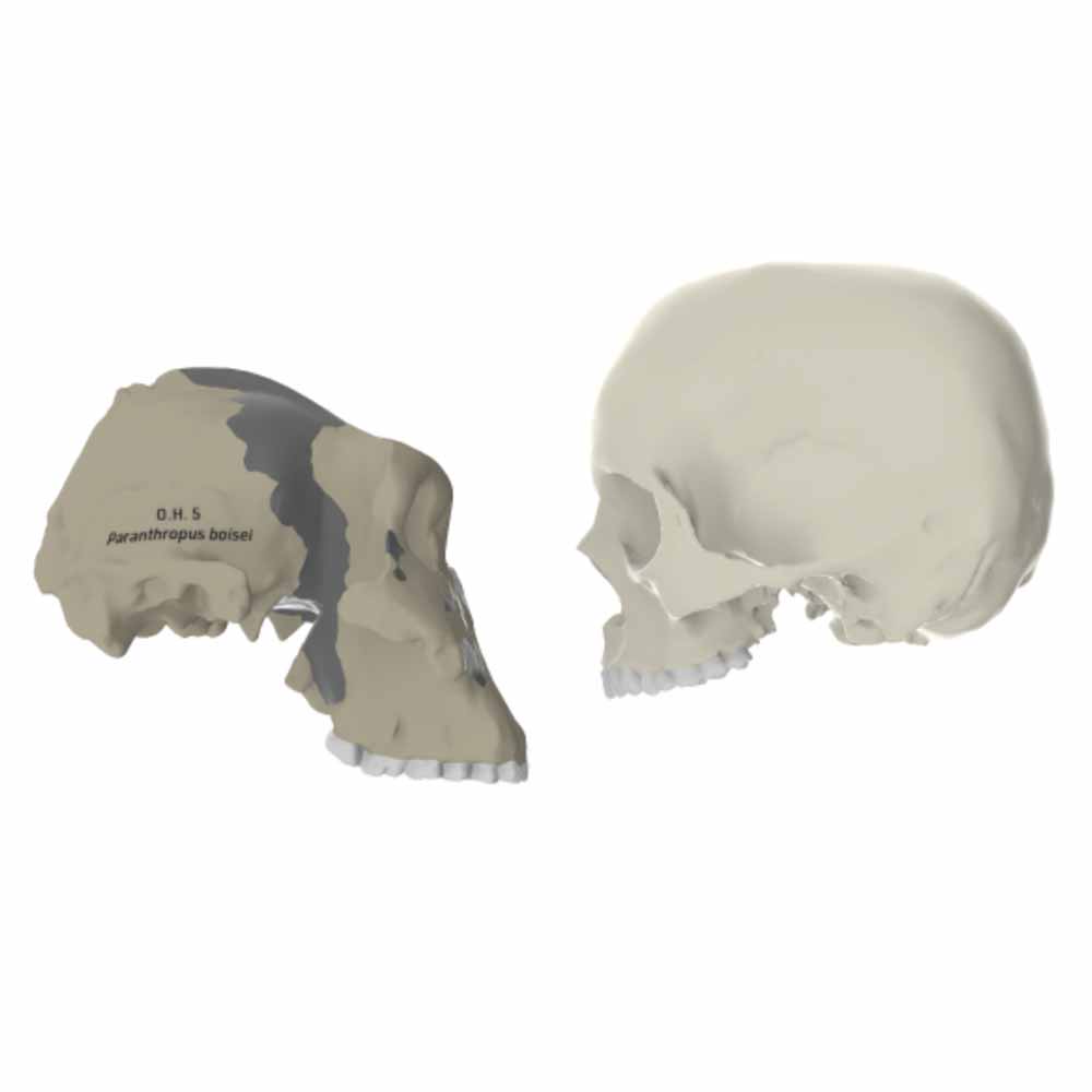
Paranthropus boisei with OH 5
The iconic skull from Olduvai Gorge compared with a modern human and Sts 5.
Homo
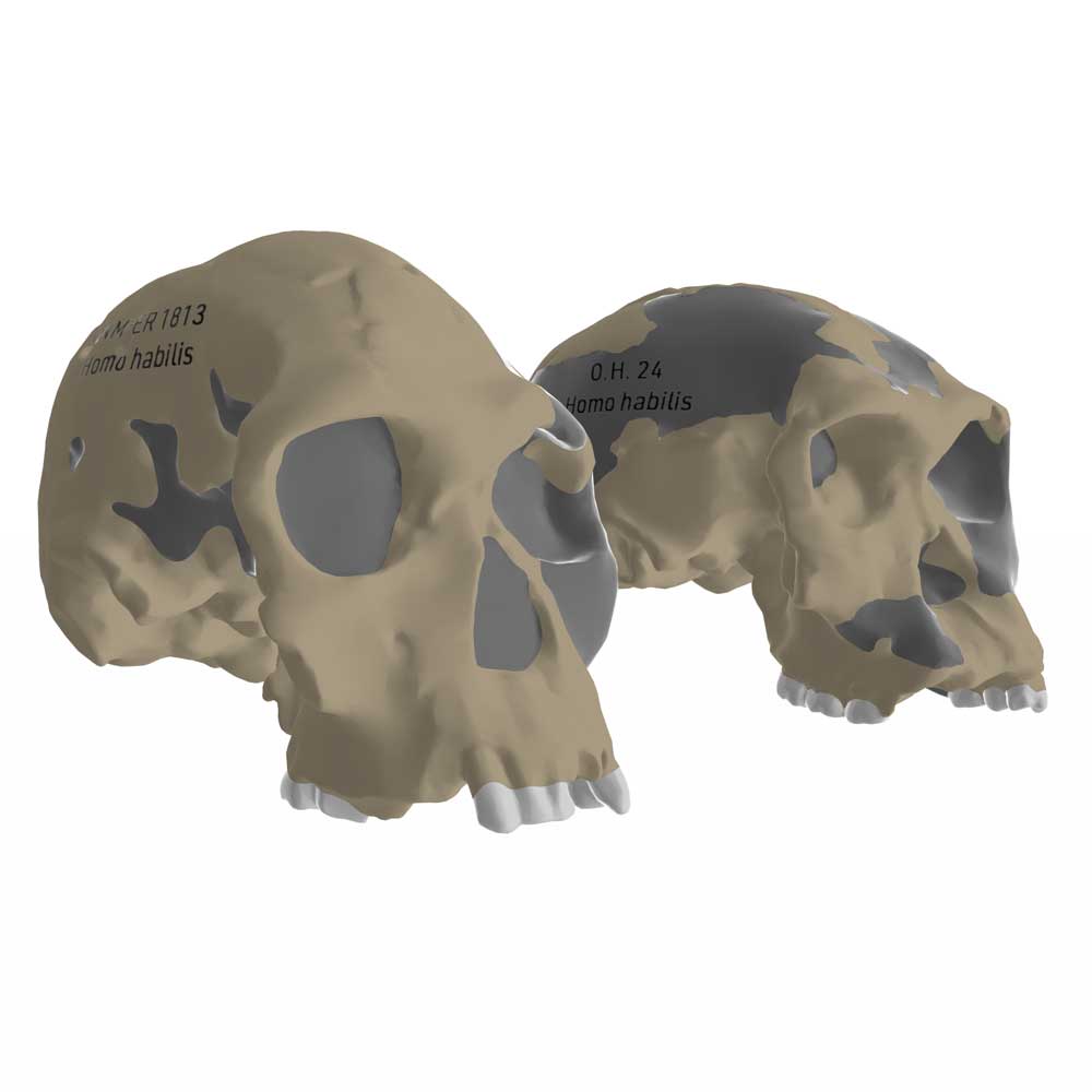
Cranial anatomy of Homo habilis
KNM-ER 1813 and OH 24 compared with modern human and Sts 5.
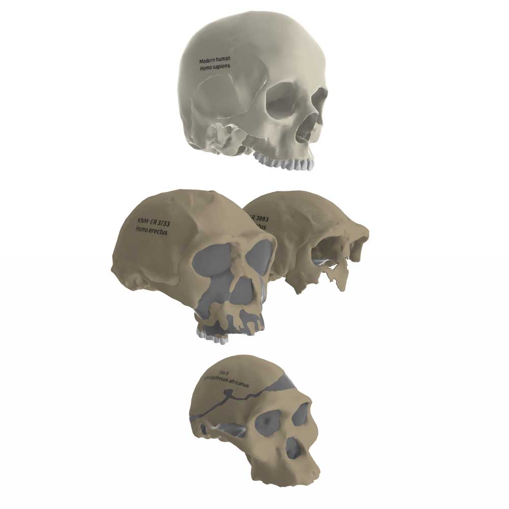
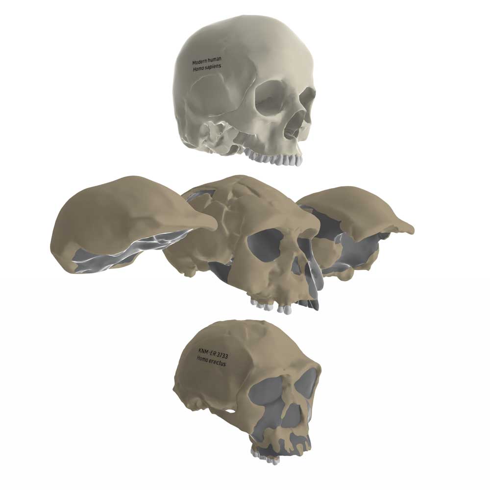
African Homo erectus crania
KNM-ER 3733 and KNM-ER 3883 as representatives of early H. erectus in Africa.
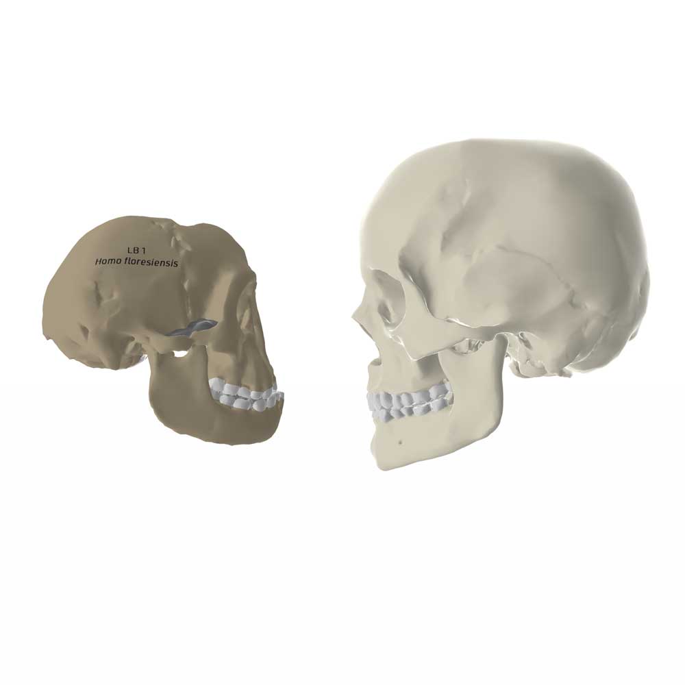
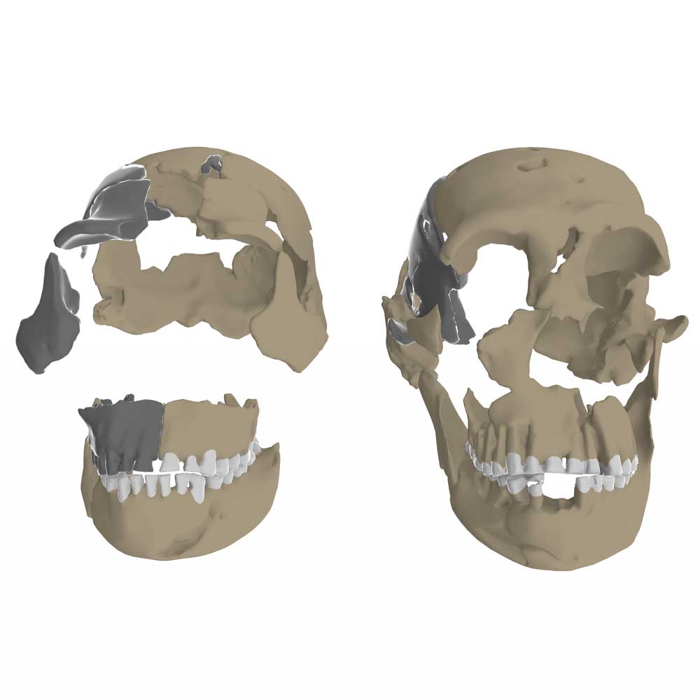
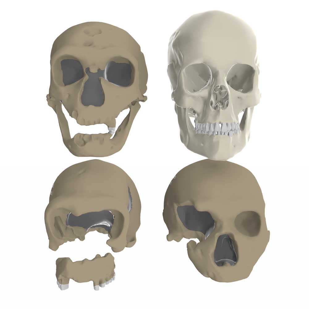
Homo floresiensis cranial anatomy
A look at the skull of the LB1 skeleton, compared to modern human and Homo erectus skulls.
Indonesian Homo erectus crania
Cranial remains from Trinil, Sangiran, and Ngandong in comparison to modern human and African H. erectus.
Cranial anatomy of Homo naledi
Three adult cranial specimens of the recently-discovered fossil hominin species.
Three Neandertal crania
Skulls from La Chapelle-aux-Saints, La Quina, and Monte Circeo illustrate Neandertal features.