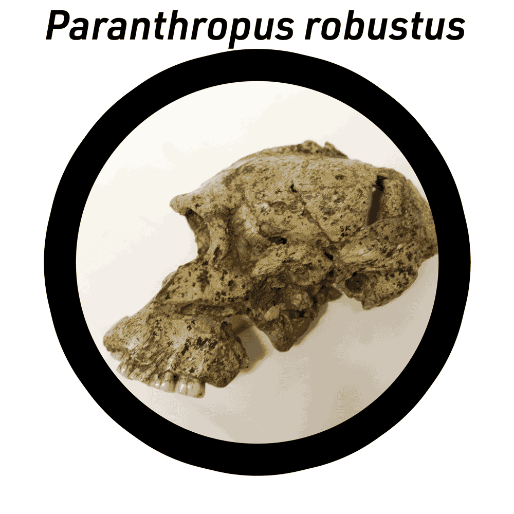
Virtual lab: Paranthropus robustus crania
Anthropologists have found Paranthropus robustus fossils at several sites in South Africa, including Kromdraai, Swartkrans, Drimolen, and Cooper's Cave. The word “robust” refers to size and strength. The body size of P. robustus was not exceptionally large, but they had large molar and premolar teeth, and a very large and thick mandible, or jawbone. The main muscles of the jaw, the temporalis muscles, were so large that they ran up the complete height of the skull to meet at the midline. The high ridge of bone where these muscles attached to the top of the skull is called the sagittal crest.

P. robustus is one of the best-represented species of early hominins. The first specimen to be found was TM 1517, a partial skeleton with cranial remains from Kromdraai, presently in the Cradle of Humankind World Heritage Site. The largest sample of P. robustus fossils come from Swartkrans, less than 3 km from Kromdraai. The iconic skull, SK 48, provides a good illustration of the anatomy of the cranium of P. robustus with its sagittal crest, large, thick cheekbones, and relatively large molar teeth.
This virtual lab includes two crania of P. robustus, TM 1517 and SK 48. The lab also includes two crania from other species to serve as a comparison, these are Sts 5, which is an example of Au. africanus from Sterkfontein, and a cranium of a modern human.
As you examine the P. robustus cranial fossils, work on the following:
- Identify the features that characterize the skulls of this species, including the sagittal crest, the postorbital constriction,
- Which aspect of each fossil is most different from the modern human skull?
- What features of the H. naledi crania are shared with Homo?
Materials in this lab
- All fossil material in this lab is curated at the Ditsong Museum of Natural History, Pretoria, South Africa. This includes the TM 1517 cranial remains, SK 48, and Sts 5. The models in this virtual lab are based upon 3D surface scans of casts in the Biological Anthropology collection of UW-Madison.
- The model of the human calvaria is based on an anatomical model created by Hannah Newey. The model is available on Sketchfab with a Creative Commons Non-Commercial Share-alike (CC-BY-NC-SA) license. I reduced the polygon count of the model for this virtual lab.
Back to full list of virtual labs