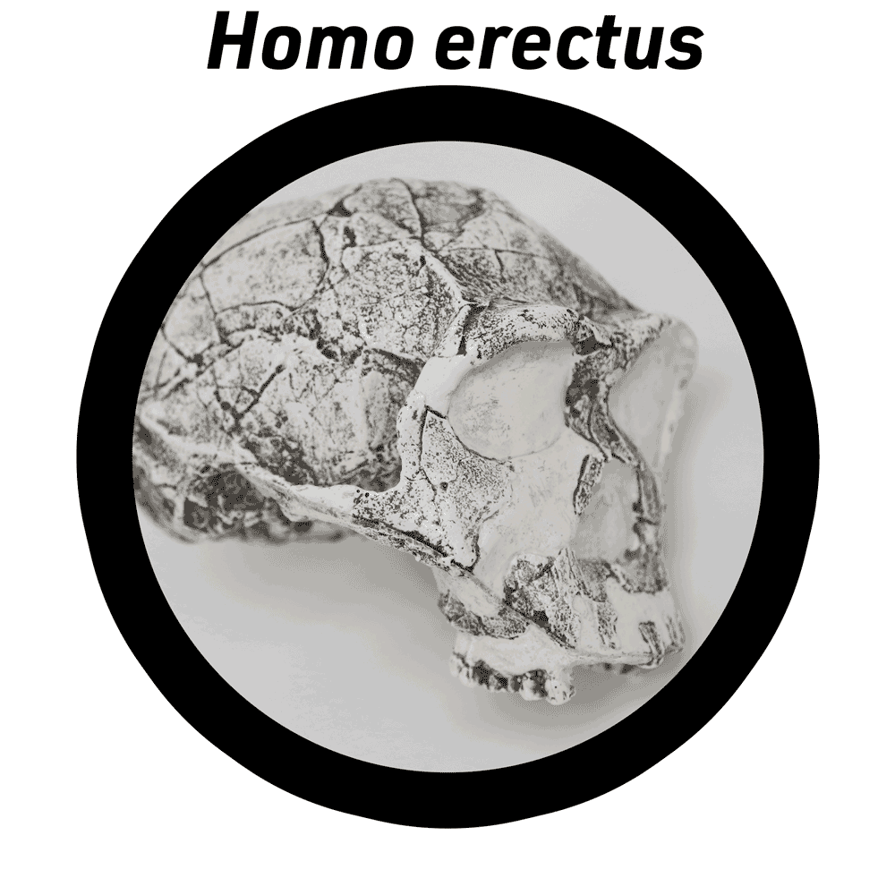
Virtual lab: Indonesian Homo erectus crania
The first discovery of Homo erectus was from the Trinil site in present-day Indonesia, in 1891. Since then, many fossils of this species have been unearthed in the surrounding region. These fossils span the period from 1.5 million years ago up to around 110,000 years ago. The latest fossils attributed to H. erectus occur in Java, where they persisted longer than anywhere else yet known.

The connection between Indonesian H. erectus populations and those in other parts of the world, including Africa and China, is not clear. The fossils from these different regions share many features of the skull. They tend to have thick cranial bone. They also share several thickened or reinforced areas of the skull, including:
- Supraorbital torus. This is present in all fossil crania of H. erectus, and is thick and projecting in many of them.
- Nuchal torus. A thickened bar of bone above the attachment area of the neck muscles, on the back of the cranium. More or less thick and projecting in different individual fossils, it is present in nearly all H. erectus adults.
- Angular torus. A thickened area associated with the posteriormost attachment of the temporalis muscle, on the parietal bone.
- Sagittal keel. H. erectus crania often have flattened areas on either side of the midline, forming an angle along the top of the skull. This can be on the frontal bone, on the parietals, or both.
Still, there are many differences among the skulls from these different parts of the world, variation within each of the regions, and differences between earlier and later skulls.
This virtual lab includes three crania of H. erectus from Indonesia, together with a cranium of a modern human and the KNM-ER 3733 fossil cranium of H. erectus from Kenya. The three Indonesian H. erectus fossils are Trinil 2, Sangiran 17, and Ngandong 10. These span a range of ages. Trinil 2 is the oldest here, estimated to be around 900,000 years old. Sangiran 17 dates to around 800,000 years ago, and the Ngandong 10 skull is between 136,000 and 106,000 years ago.
These fossil H. erectus crania share many features but also exhibit some differences that may reflect their evolution. As you examine these skulls consider the following:
- Outline the features of H. erectus discussed above on these crania.
- Consider the skulls in order of their geological age. Are there features that seem to change over time in the sample?
- Genetic evidence from recent people in island Southeast Asia and New Guinea suggests that a population known as the Denisovans make up a small amount of the ancestry of these living people. These Denisovans share a common origin with the Neandertals around 600,000 years ago, and their common heritage with African ancestors of living people is more recent than the Trinil or Sangiran skulls. Looking at the Ngandong 10 skull, could it represent a different population from the earlier Sangiran and Trinil cranial remains?
Materials in this lab
- The original Trinil 2 fossil is curated at the Naturalis Museum in Leiden, Netherlands. The model in this virtual lab draws upon data from a cast in the Biological Anthropology collection at UW-Madison.
- The original Sangiran 17 fossil cranium is curated at the Geological Research and Development Centre, Bandung, Indonesia. The model in this virtual lab is based upon measurements and photographs of casts and the original specimen.
- The Ngandong 10 specimen is curated at Gadjah Mada University in Yogyakarta, Indonesia. The model in this virtual lab is based upon data from a cast in the Biological Anthropology collection at UW-Madison.
- The original KNM-ER 3733 cranium is curated at the Nairobi National Museum, Nairobi, Kenya. The model in this laboratory are based upon data from a cast in the Biological Anthropology collection at UW-Madison. A high-quality 3D scan of this fossil is available from AfricanFossils.org. That model is compatible with 3D printing and classroom use.
- The model of the human calvaria is based on an anatomical model created by Hannah Newey. The model is available on Sketchfab with a Creative Commons Non-Commercial Share-alike (CC-BY-NC-SA) license. I reduced the polygon count of the model for this virtual lab.
Back to full list of virtual labs