The cranium is a structure composed of 28 separate bones in most people. Six of these are the bones of the middle ear on left and right sides, and one is the mandible. The remaining 21 bones are fused together in adults at immobile joints known as sutures.
The teeth are rooted in the mandible and in the left and right maxillary bones.
Some bones of the skull are paired bones, with both a left and a right sided bone that mirror each other. When anthropologists refer to these bones, they always include the side, left or right. Other bones are unpaired bones, which lie on the midline of the skull and are symmetrical in form with left and right halves.
Basic divisions of the cranium
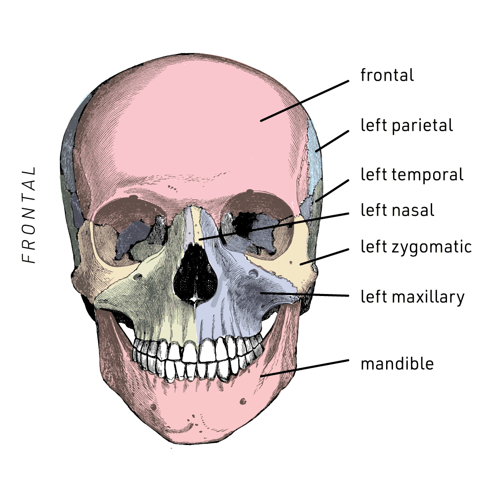
Cranial bones, as visible in anterior view. Image: John Hawks, based on Quain's Anatomy
The calvaria includes all of the cranial bones except the mandible.
The neurocranium, often called the cranial vault, is made up of the bones that enclose the brain.
The viscerocranium is the skeleton of the face, including the mandible.
Bones of the neurocranium
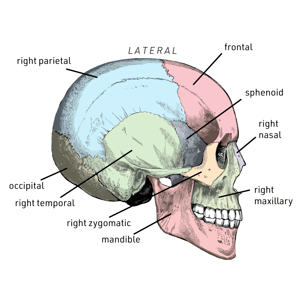
Cranial bones, as visible in right lateral view. Image: John Hawks, based on Quain's Anatomy
Frontal bone: The bone of the forehead, including the superior portion of the orbits, or eye sockets.
Left and right parietal bones: These two bones together make up much of the top and sides of the vault.
Left and right temporal bones: The lower part of the sides of the vault, including the ear opening, or external acoustic porus.
Sphenoid bone: Just anterior to the temporal bones, the sphenoid is a single bone that stretches through the skull from left to right.
Occipital bone: This bone is the posterior wall of the vault and is the largest bone of the cranial base. The foramen magnum, which accommodates the spinal cord, is a large hole in the occipital bone.
Ethmoid bone: This bone is visible in the medial walls of the orbits, where it lies inferior to the frontal bone and anterior to the sphenoid. It also makes up the superior part of the nasal septum.
Bones of the viscerocranium
Mandible: The bone of the lower jaw.
Left and right maxillary bones: The maxillary bones are the largest bones of the face. They make up part of the medial and inferior border of the orbits, the lateral border of the nasal aperture, and the alveoli for the roots of the maxillary teeth.
Left and right zygomatic bones: These are the cheekbones, which also make up the lateral and part of the inferior border of the orbit. The zygomatic arches at the sides of the skull are composed of portions of the zygomatic and temporal bones.
Left and right nasal bones: These are small bones that form the bony portion of the bridge of the nose.
Left and right lacrimal bones: These small thin bones are in the medial wall of each orbit, anterior to the ethmoid bone. Each includes a lacrimal foramen, which accommodates the tear duct.
Left and right inferior nasal conchae: Each inferior nasal concha supports membranes of the nose and attach to the corresponding (left or right) maxillary bone upon the lateral wall of the nasal aperture.
Left and right palatine bones: The palatine bones include thin plates of bone at the back of the hard palate, posterior to the left and right maxillary bones. They also include a portion that extends superiorly, making up part of the lateral wall of the nasal passages.
Vomer: This bone is a thin plate that is part of the nasal septum, meeting the ethmoid bone on its superior and anterior border.
Materials in this lab
The human cranial bone 3D models in this virtual lab have been provided under a Creative Commons license by The Database Center for Life Science, Japan. The license is CC Attribution-Share Alike 2.1 (CC-BY-SA). These are anatomical models created by digital artists based on their study of human anatomy. For this virtual lab, all models have been reduced substantially in polygon count. The original, full-resolution models can be found on the BodyParts3D website.