
Virtual lab: Morphology of the femur
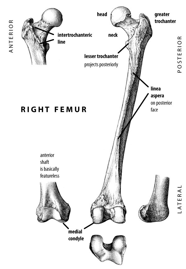
The femur is the bone of the upper leg. The proximal end of the femur connects to the hip joint. It is marked by a spherical ball, called the femoral head, that fits into the socket of the hip joint, the acetabulum. The head is connected to the shaft of the femur by an elongated segment of bone, called the femoral neck. Lateral to the neck, a large projection juts proximally off the top of the bone, called the greater trochanter. A smaller projection on the posterior surface of the femur, just below the neck, is called the lesser trochanter.
The shaft of the femur is thick, and may be quite straight or slightly curved from front to back.
The distal end of the femur connects to the knee joint. It is marked by two large articular processes, called condyles, which sit on top of the tibia.
- The human femur models in this virtual lab come from two sources. One left and one right femur have been provided under a Creative Commons license by The Database Center for Life Science, Japan. The license is CC Attribution-Share Alike 2.1 (CC-BY-SA). The original, full-resolution model can be found on the BodyParts3D website.
- The other left and right femora are based on scans of osteological specimens in the Biological Anthropology collection of UW-Madison.
Siding a femur
Telling a right femur from a left is easy if you know what to look for. The head of the femur connects to the hip joint, so it is toward the middle of the body. Using anatomical terminology, the head is medial and proximal. The greater trochanter is on the lateral side of the bone, away from the middle of the body. The front side of the femur (called the anterior side) is fairly smooth. The back side (called the posterior side) has the lesser trochanter and the condyles both projecting back.
If you align the femur so that the condyles and lesser trochanter are pointing backward, the head must point toward the hip. Also, if you place the condyles of a human femur flat on a table surface, the shaft of the bone will have an angle, called the valgus angle. It angles outward toward the hip from the center of mass of the body. So a right femur angles toward the right, a left femur angles toward the left.
Remember, right and left refer to the skeleton’s body, not the way you are looking at it!
Materials in this lab
Back to full list of virtual labs